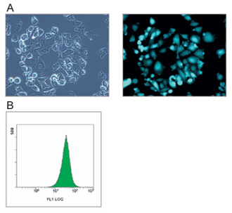
| Fig. 9 | Characterization of CHO-EGFP cell line. A) Fluorescence images of CHO-EGFP cells; left, phase contrast image (ü~400); right, fluorescence image (ü~400). CHO cells were transfected with pCMV-IE EGFP and established by a cloning assay using a 96-well plate. The constant expression of the EGFP gene in the CHO-EGFP cells was analyzed under a fluorescence microscope. B) Flow cytometry analysis of CHO-EGFP cells. The cells were gently harvested using 0.2% trypsin-EDTA solution and EGFP expression was analyzed on a flow cytometer (5.0ü~104 events, 488-nm excitation laser wavelength, 500 V). |

