Ā@
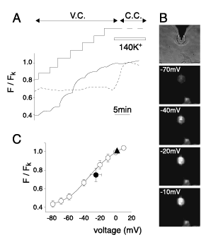
Fig.3Ā@Ā@Calibration of the relative fluorescence of DiBAC4(3) against clamp potentials. (A) Changes in relative fluorescence intensity (F/FK) were measured in standard solution at depolarization voltage steps in the range of –80 and +10 mV under voltage-clamp (solid line) in HEKBKÉŅÉņ stained with 100 nmol/l DiBAC4(3). V.C. and C.C. indicate voltage-clamp and current clamp, respectively. The F/FK in not voltage-clamped cells was shown in the same time course by broken line. Single cells were selected for the recording based on cell capacitance (<30 pF). At the end of the procedure, FK was measured under current-clamp mode in 140 mmol/l K+ solution as indicated by horizontal bar. (B) Transmitted images of cells and a pipette and fluorescence images of cells, which were voltage clamped or not clamped. The cell attached by the pipette was voltage clamped. F intensity in the voltage-clamped cell depended on the clamp potentials, but that in non-clamped cell did not. The relationships between F/FK and clamp potentials and non-clamped cell are shown in "A". (C) The relationship between F/FK and corresponding clamp potentials. The solid line represents the theoretical fit to the data points using the Boltzmann equation (n=5). F/Fk=1.1-0.7/{1+exp[(E+31)/17.8]},where E is the membrane potential(abscissa). Closed triangle (n=5) and closed circle (n=5) represent the results in 140mmol/l K+ solution and the standard solution under current-clamp mode, respectively. Reference 16) with the permission from Japanese Pharmacological Society.

