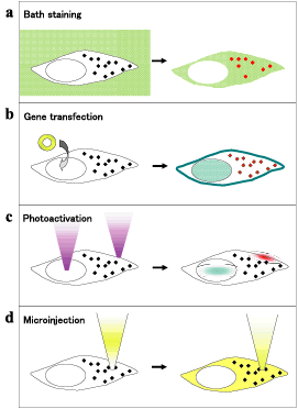
Fig.3
Live cell fluorescent marking by 4 ways (a-d). Left images were before loading of fluorophores, and right were after the staining.