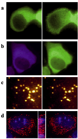
| Fig.2 Spatio-temporal analysis for live cell functions and movements. a, translocation of PKC-GFP. b, Ca ions by FRET between CFP and YFP (Yellow Cameleon 2.1). c, exocytosis of acridine orange containing granules. d, multi-angle views of mitochondria (DsRed as red) and nucleus (CFP as blue). In a-c, left images were before stimulation (resting states), and right were after stimulation. All figures were recorded at 12th Medical Photonics Course, Hamamatsu, 2003. |

