|
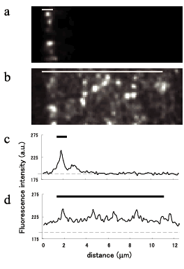
Fig. 2 Imaging of tetramethylrhodamine (TMR) molecules in different widths of illumination field. Horizontal bars indicate the position of illumination, and the width is set approximately at 1 (a & c) and 10 (b & d) mm. Each bright spot (in a & b) indicates a single molecule of TMR. Horizontal dotted line in line profiles (c, d), dark level of video camera. |

